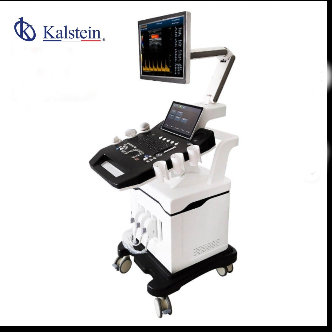
Color Doppler Ultrasound Cart with Wheels 3D Model YR05149
Color Doppler Ultrasound Cart with Wheels 3D Model YR05149 — Kalstein laboratory equipment with technical specifications, advanced features, and certified professional solutions for scientific use.
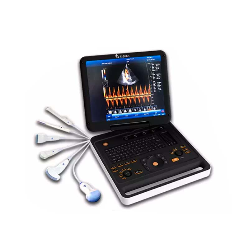
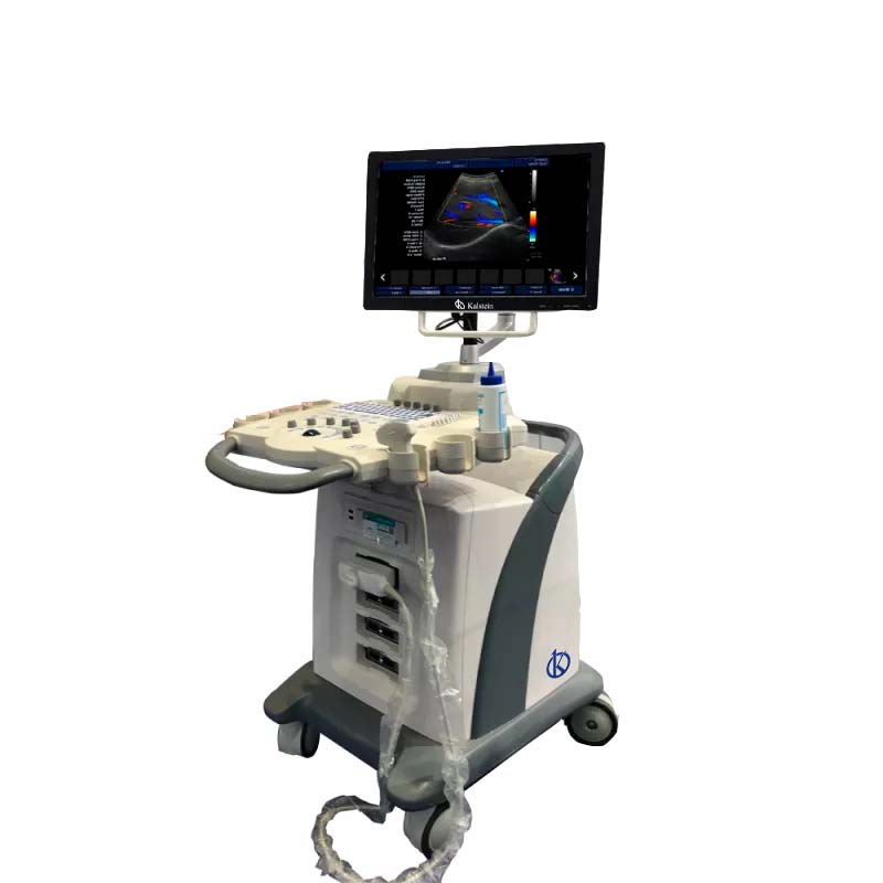
The Color Doppler Ultrasound Cart with Wheels 3D Model YR05149 is a state-of-the-art diagnostic tool designed for precision and convenience in medical imaging. Featuring a 15-inch LED screen with multilingual capabilities, this ultrasound cart supports a range of imaging technologies including CF+B mode, PDI, DPDI, TDI, and TSI. The device also allows for trapezoidal imaging, virtual convex matrix, and matrix expansion, ensuring versatility for various diagnostic needs. With advanced features like automatic IMT measurement, endometrial assessment, and quality control, this model is an indispensable asset in fields such as obstetrics and cardiology. The built-in DICOM 3.0 protocol facilitates seamless printing and data sharing, while the integration of workstations and databases enhances workflow efficiency. Enjoy the added convenience of a portable design with wheels, making it perfect for dynamic healthcare environments. Upgrade your practice with this comprehensive diagnostic solution, and take advantage of our platform Kalstein Plus to request a customized quote today.
Market Price
The Color Doppler Ultrasound Cart with Wheels 3D Model YR05149 typically ranges from $7500 to $8300 USD in the market. This price range is reflective of the advanced technology and versatility this equipment offers, making it a valuable investment for medical facilities.
Frequently Asked Questions
How many transducer ports are available? This model comes with 4 transducer ports, allowing simultaneous connections for varied diagnostic procedures.
Can the ultrasound cart connect to external devices? Yes, it offers multiple connectivity options, including USB, Ethernet, VGA, and HDMI ports for enhanced compatibility with external devices.
Advantages and Disadvantages
Advantages: The ultrasound cart is highly versatile and portable, with advanced imaging techniques and user-friendly features. It supports a wide range of diagnostic applications with ease.
Disadvantages: The initial investment may be high for smaller clinics, but the long-term benefits and efficiency can offset the cost.
Product Use in the Field
This ultrasound cart is ideal for various clinical environments, particularly in obstetrics and cardiology, where precise and versatile imaging solutions are necessary. It offers quick transitions between imaging modes and supports comprehensive diagnostic assessments, enhancing patient care.
Recommendations
To maximize the lifespan and performance of the ultrasound cart, conduct regular maintenance checks and ensure software updates are applied timely. Proper operator training is recommended to utilize all functions effectively and improve diagnostic outcomes.
Features
- 15″ LED screen with multilingual function
- Model CF+B simultaneous imaging, PDI, DPDI, TDI, TSI support
- Automatic IMT and endometrial measurements
- Seamless DICOM 3.0 protocol integration
- Efficient workflow with integrated workstations and databases
- User-friendly online guidance and control
Technical Specifications
| Model | YR05149 | |||
| Connectivity/Media/Peripherals | ||||
| Transducer Ports | 4 | |||
| USB ports | 4 | |||
| HDD | 64GB (SSD), 120G/200GB SSD (optional) | |||
| foot switch | USB | |||
| Ethernet port | 2(100Mb/1000Mb) | |||
| External screen | VGA,HDMI, | |||
| Printer (Optional) | USB Printer, Digital Laser Printer, B/W Digital Thermal Printer | |||
| Printing area | Image, report, Image+report | |||
| Cine/Picture Memory | ||||
| Memory Cinema | 1200 frames (max) | |||
| Film Review Speed | 1, 2, 4, 8 | |||
| Cinema Review Loop | YES | |||
| Capture function | YES | |||
| DICOM connectivity | DICOM3.0 Compliant | |||
| 3Dsoftware | Built-in 3D software | |||
| Image Storage | Storage Format: PNG, AVI, BMP, JPEG, DICOM Export Video Format: AVI Export Image Format: PNG, JPEG, BMP, DICOM USB Flash Drive | |||
| Technology | ||||
| Panoramic Imaging Technology Full Digital Signal Processing Technology Multi-Beamforming Technology Spot Reduction Technology Tissue Harmonic Imaging Technology Dynamic Tissue Optimization Technology Duplex and Triplex Synchronous Display Directional Power Doppler Imaging Parameters Imaging Parameters special tissue presets PW Auto Trace Update online CF+B mode on one screen Complex model imaging Automatic IMT measurements Virtual Convex Array Trapezoidal imaging | ||||
| Overall Performance | ||||
| Digital Broadband | 12288 channels | |||
| Beam Former | reprogrammable | |||
| Transmission voltage | Adjustable (15 steps) | |||
| Beamformer Frequency Range | 1~40MHz | |||
| Led monitor | ||||
| Size (diagonal) | fifteen" | |||
| Contrast Ratio | 800: 01: 00 | |||
| Resolution | 1024×768 pixels | |||
| Brightness | 230 cd / m2 | |||
| Color Depth | 24 bit | |||
| Rotation Angle | ±90° | |||
| Gray levels | 256 | |||
| Imaging performance | ||||
| Start Time (max.) | Average < 90 seconds | |||
| Preset Switching Time | Average < 1 second | |||
| Storage Time (Image to disk) | Average < 0.5 seconds | |||
| transducers | ||||
| Research | Convex array probe | Linear Array Probe | intracavitary probe | microconvex probe |
| Frequency | Center 3.5MHz | Central 7.5MHz | Center 6.5MHz | Center 4.0MHz |
| (2.0MHz to 10.0MHz) | (6.0MHz to 10.0MHz) | (5.0MHz to 9.0MHz) | (2.0MHz to 5.5MHz) | |
| field of play | 0.516mm | 0.352mm | 0.216mm | |
| Radio | 60mm | N/A | 10mm | |
Technical specifications
| Dimensions | L 162 × W 72 × H 95 cm |
|---|---|
| Weight | 222 kg |
| Manufacturer | Kalstein |
Video Color Doppler Ultrasound Cart with Wheels 3D Model YR05149
Frequently asked questions
How to know the prices of Color Doppler Ultrasound Cart with Wheels 3D Model YR05149?
To know the price of Color Doppler Ultrasound Cart with Wheels 3D Model YR05149, please send us an email with your request through the contact form AQUI.
What are the delivery times for Color Doppler Ultrasound Cart with Wheels 3D Model YR05149?
Delivery time depends on stock availability and freight type (air or sea). In stock: air 15-30 days, sea 45-60 days. Out of stock: air 30-60 days, sea 60-90 days.
How to make a purchase of Color Doppler Ultrasound Cart with Wheels 3D Model YR05149?
You can buy by email ([email protected]), phone (+33 (0) 1 70 39 26 50) or through the official Kalstein website in your country.
How does the warranty work for Color Doppler Ultrasound Cart with Wheels 3D Model YR05149?
All Kalstein equipment comes with a 1-year warranty against manufacturing defects. The warranty does not cover damage from improper installation or misuse. See our «terms and conditions» AQUI.
Can I request a quote online for Color Doppler Ultrasound Cart with Wheels 3D Model YR05149?
Yes, you can request a quote for the Kalstein equipment you are interested in directly from our official website. Click AQUI.